Pages

Monday, May 1, 2017

Evidence of a 'higher' state of consciousness


Evidence of a 'higher' state of consciousness


Scientific evidence of a 'higher' state of consciousness has been found in a new study. Using brain imaging technology, researchers measured the tiny magnetic fields produced in the brain and found that, across three psychedelic drugs, one measure of conscious level -- the neural signal diversity -- was reliably higher.

Image created using brain imaging technology, showing changes in neural signal diversity while under the influence of LSD.

Scientific evidence of a 'higher' state of consciousness has been found in a study led by the University of Sussex.
Neuroscientists observed a sustained increase in neural signal diversity -- a measure of the complexity of brain activity -- of people under the influence of psychedelic drugs, compared with when they were in a normal waking state.
The diversity of brain signals provides a mathematical index of the level of consciousness. For example, people who are awake have been shown to have more diverse neural activity using this scale than those who are asleep.
This, however, is the first study to show brain-signal diversity that is higher than baseline, that is higher than in someone who is simply 'awake and aware'. Previous studies have tended to focus on lowered states of consciousness, such as sleep, anaesthesia, or the so-called 'vegetative' state.
The team say that more research is needed using more sophisticated and varied models to confirm the results but they are cautiously excited.
Professor Anil Seth, Co-Director of the Sackler Centre for Consciousness Science at the University of Sussex, said: "This finding shows that the brain-on-psychedelics behaves very differently from normal.
"During the psychedelic state, the electrical activity of the brain is less predictable and less 'integrated' than during normal conscious wakefulness -- as measured by 'global signal diversity'.
"Since this measure has already shown its value as a measure of 'conscious level', we can say that the psychedelic state appears as a higher 'level' of consciousness than normal -- but only with respect to this specific mathematical measure."
For the study, Michael Schartner, Adam Barrett and Professor Seth of the Sackler Centre reanalysed data that had previously been collected by Imperial College London and the University of Cardiff in which healthy volunteers were given one of three drugs known to induce a psychedelic state: psilocybin, ketamine and LSD.
Using brain imaging technology, they measured the tiny magnetic fields produced in the brain and found that, across all three drugs, this measure of conscious level -- the neural signal diversity -- was reliably higher.
This does not mean that the psychedelic state is a 'better' or more desirable state of consciousness, the researchers stress; instead, it shows that the psychedelic brain state is distinctive and can be related to other global changes in conscious level (e.g. sleep, anaesthesia) by application of a simple mathematical measure of signal diversity. Dr Muthukumaraswamy who was involved in all three initial studies commented: "That similar changes in signal diversity were found for all three drugs, despite their quite different pharmacology, is both very striking and also reassuring that the results are robust and repeatable."
The findings could help inform discussions gathering momentum about the carefully-controlled medical use of such drugs, for example in treating severe depression.
Dr Robin Cahart-Harris of Imperial College London said: "Rigorous research into psychedelics is gaining increasing attention, not least because of the therapeutic potential that these drugs may have when used sensibly and under medical supervision.
"The present study's findings help us understand what happens in people's brains when they experience an expansion of their consciousness under psychedelics. People often say they experience insight under these drugs -- and when this occurs in a therapeutic context, it can predict positive outcomes. The present findings may help us understand how this can happen."
As well as helping to inform possible medical applications, the study adds to a growing scientific understanding of how conscious level (how conscious one is) and conscious content (what one is conscious of) are related to each other.
Professor Seth said: "We found correlations between the intensity of the psychedelic experience, as reported by volunteers, and changes in signal diversity. This suggests that our measure has close links not only to global brain changes induced by the drugs, but to those aspects of brain dynamics that underlie specific aspects of conscious experience."
The research team are now working hard to identify how specific changes in information flow in the brain underlie specific aspects of psychedelic experience, like hallucinations.

New eye test detects earliest signs of glaucoma


New eye test detects earliest signs of glaucoma


Researchers have developed a simple, inexpensive diagnostic tool DARC (Detection of Apoptosing Retinal Cells). In clinical trials it allowed for the first time visualization of individual nerve cell death in patients with glaucoma. Early detection means doctors can start treatments before sight loss begins. Ongoing trials are investigating the potential of the test for other neurodegenerative conditions.


A simple eye test could help solve the biggest global cause of irreversible blindness, glaucoma.
In clinical trials, the pioneering diagnostic -- developed by researchers at University College London (UCL) and the Western Eye Hospital -- allowed doctors to see individual nerve cell death in the back of the eye.
Glaucoma affects 60 million people in the world, with 1 in 10 suffering total sight loss in both eyes.
Early detection means doctors can start treatments before sight loss begins. The test also has potential for early diagnosis of other degenerative neurological conditions, including Parkinson's, Alzheimer's and multiple sclerosis.
Results of first clinical trials with glaucoma patients are published in the journal BRAIN.
Professor Francesca Cordeiro at UCL Institute of Ophthalmology, who led the research, said: "Detecting glaucoma early is vital as symptoms are not always obvious. Although detection has been improving, most patients have lost a third of vision by the time they are diagnosed. Now, for the first time, we have been able to show individual cell death and detect the earliest signs of glaucoma. While we cannot cure the disease, our test means treatment can start before symptoms begin. In the future, the test could also be used to diagnose other neurodegenerative diseases."
Loss of sight in patients with glaucoma is caused by the death of cells in the retina at the back of the eye. This cell death is called apoptosis.
As with other neurodegenerative conditions, more and more nerve cells are lost as the disease progresses.
Professor Philip Bloom, Chief Investigator at Western Eye Hospital, part of Imperial College Healthcare NHS Trust, added: "Treatment is much more successful when it is begun in early stages of the disease, when sight loss is minimal. Our developments mean we could diagnose patients 10 years earlier than was previously possible."
The technique developed is called DARC, which stands for detection of apoptosing retinal cells. It uses a specially developed fluorescent marker which attaches to cell proteins when injected into patients. Sick cells appear as white fluorescent spots during eye examination. UCL Business, the commercialisation company of UCL, holds the patents for the technology.
The examination uses equipment used during routine hospital eye examinations. Researchers hope that eventually it may be possible for opticians to do the tests, enabling even earlier detection of the disease.
Bethan Hughes, from Wellcome's Innovation team said: "This innovation has the potential to transform lives for those who suffer loss of sight through glaucoma, and offers hope of a breakthrough in early diagnosis of other neurodegenerative diseases. Loss of sight as you age is an incredibly difficult disability, impacting quality of life and independence."
Initial clinical trials were carried out on a small number of glaucoma patients and compared with tests on healthy people. The initial clinical trials established the safety of the test for patients.
Further studies will now be carried out to into DARC and how it can be used not only to diagnose and treat glaucoma patients but also for other neurodegenerative conditions.

When students pay attention in class, their brains are in sync


When students pay attention in class, their brains are in sync


When people in a group are engaged with each other and with the world around them, their brainwaves show similar patterns. That's the conclusion of researchers who used portable EEG to simultaneously record brain activity from a class of high school students over the course of a semester as they went about their classroom activities. The findings highlight the promise of investigating the neuroscience of group interactions in real-world settings.


When people in a group are more engaged with each other and with the world around them, their brainwaves show remarkably similar patterns. That's the conclusion of researchers who used portable EEG to simultaneously record brain activity from an entire class of high school students over the course of a full semester as they went about their regular classroom activities. The findings reported in Current Biology on April 27 highlight the promise of investigating the neuroscience of group interactions in real-world settings.
"We found that students' brainwaves were more in sync with each other when they were more engaged during class," says co-lead author Suzanne Dikker of New York University and Utrecht University. "Brain-to-brain synchrony also reflected how much students liked the teacher and how much they liked each other. Brain synchrony was also affected by face-to-face social interaction and students' personalities. We think that all these effects can be explained by shared attention mechanisms during dynamic group interactions."
The researchers, led by David Poeppel of New York University and the Max Planck Institute of Empirical Aesthetics, used portable EEG to simultaneously record the students' brain activity. Researchers Lu Wan and Mingzhou Ding of the University of Florida then used novel analyses to assess the extent to which that brain activity was synchronized across students and how the degree of synchrony varied with class engagement and social dynamics.
The researchers think that the level of synchrony comes from a well-known phenomenon called neural entrainment. "Your brainwaves 'ride' on top of the sound waves or light patterns in the outside world, and the more you pay attention to these temporal patterns, the more your brain locks to those patterns," Dikker explains. "So, if you and the person next to you are more engaged, your brainwaves will be more similar because they are locking onto the same information."
Brain synchrony most likely supports synchronized behavior during human interaction. For example, synchrony is required for two or more people to have a good conversation, walk down the street, or dance, or carry a heavy piece of furniture. The findings suggest that social dynamics matter, even when people are just listening to the same lecture or watching the same video.
The researchers are now designing large-scale projects in which they'll be able to record brain data and other biometrics from up to 45 people simultaneously in an auditorium. They hope to answer questions such as, "What are the 'optimal' conditions for an audience to experience a performance or movie? Is there an ideal group size? Does having some joint interaction right before a performance improve the experience? How does the audience affect the performer and vice versa?"

Compact fiber optic apparatus shines light on breath analysis in real-time


Compact fiber optic apparatus shines light on breath analysis in real-time


An affordable gas sensor monitors trace levels of health-indicating chemicals, paving the way for future non-invasive studies, describe researchers in a new report.

Optical setup for gas analysis.

Affordable gas sensor setup developed by Tohoku University team monitors trace levels of health-indicating chemicals, paving the way for future non-invasive studies.
Using hollow-core optical fibre as a sensitive gas cell, researchers in Japan have devised a relatively simple and affordable sensor for monitoring biomarkers in human breath at low concentrations. Trace amounts of gases exhaled through the nose and mouth offer clues to respiratory conditions such as asthma, as well as other easy-to-administer health screening opportunities.
Tohoku University scientists explained how their device works in the journal Sensors, using isoprene as an example. Isoprene is a known indicator of cholesterol synthesis and could offer valuable insights into a patient's metabolism. Breath measurements taken from volunteers throughout the day allowed researchers to track changes in isoprene levels following activities such as exercise or eating.
Breath analysis has attracted much attention because it is non-invasive and has the potential to inform users on a range of health topics. However, detecting biomarkers present at low-concentrations often requires bulky and expensive laboratory systems. The Japanese scientists believe their sensor paves the way for a more portable and affordable solution.
Key to the success of the group's apparatus is a 3-meter-long, hollow-core optical fibre, which is coated on the inside with a reflective film. Participants breathe into a connecting tube that guides the exhaled gases into the fibre's core, where the contents are exposed to laser-driven ultraviolet light. A detector placed at the end of the optical path highlights any portions of the light signal that are absorbed as the ultraviolet emission passes through the gas sample. And this series of so-called 'absorption peaks' forms a chemical signature that reveal which molecules are present. The combination of a long beam-path and high-intensity emission enhances the measurement, so even chemicals present at parts-per-billion levels can be detected.

Stem cells edited to fight arthritis


Stem cells edited to fight arthritis


Using CRISPR technology, a team of researchers rewired stem cells' genetic circuits to produce an anti-inflammatory arthritis drug when the cells encounter inflammation. The technique eventually could act as a vaccine for arthritis and other chronic conditions.

Scientists have rewired stem cells' genetic circuits to produce an anti-inflammatory arthritis drug when the cells encounter inflammation.

Using new gene-editing technology, researchers have rewired mouse stem cells to fight inflammation caused by arthritis and other chronic conditions. Such stem cells, known as SMART cells (Stem cells Modified for Autonomous Regenerative Therapy), develop into cartilage cells that produce a biologic anti-inflammatory drug that, ideally, will replace arthritic cartilage and simultaneously protect joints and other tissues from damage that occurs with chronic inflammation.
The cells were developed at Washington University School of Medicine in St. Louis and Shriners Hospitals for Children-St. Louis, in collaboration with investigators at Duke University and Cytex Therapeutics Inc., both in Durham, N.C. The researchers initially worked with skin cells taken from the tails of mice and converted those cells into stem cells. Then, using the gene-editing tool CRISPR in cells grown in culture, they removed a key gene in the inflammatory process and replaced it with a gene that releases a biologic drug that combats inflammation.
The research is available online April 27 in the journal Stem Cell Reports.
"Our goal is to package the rewired stem cells as a vaccine for arthritis, which would deliver an anti-inflammatory drug to an arthritic joint but only when it is needed," said Farshid Guilak, PhD, the paper's senior author and a professor of orthopedic surgery at Washington University School of Medicine. "To do this, we needed to create a 'smart' cell."
Many current drugs used to treat arthritis -- including Enbrel, Humira and Remicade -- attack an inflammation-promoting molecule called tumor necrosis factor-alpha (TNF-alpha). But the problem with these drugs is that they are given systemically rather than targeted to joints. As a result, they interfere with the immune system throughout the body and can make patients susceptible to side effects such as infections.
"We want to use our gene-editing technology as a way to deliver targeted therapy in response to localized inflammation in a joint, as opposed to current drug therapies that can interfere with the inflammatory response through the entire body," said Guilak, also a professor of developmental biology and of biomedical engineering and co-director of Washington University's Center of Regenerative Medicine. "If this strategy proves to be successful, the engineered cells only would block inflammation when inflammatory signals are released, such as during an arthritic flare in that joint."
As part of the study, Guilak and his colleagues grew mouse stem cells in a test tube and then used CRISPR technology to replace a critical mediator of inflammation with a TNF-alpha inhibitor.
"Exploiting tools from synthetic biology, we found we could re-code the program that stem cells use to orchestrate their response to inflammation," said Jonathan Brunger, PhD, the paper's first author and a postdoctoral fellow in cellular and molecular pharmacology at the University of California, San Francisco.
Over the course of a few days, the team directed the modified stem cells to grow into cartilage cells and produce cartilage tissue. Further experiments by the team showed that the engineered cartilage was protected from inflammation.
"We hijacked an inflammatory pathway to create cells that produced a protective drug," Brunger said.
The researchers also encoded the stem/cartilage cells with genes that made the cells light up when responding to inflammation, so the scientists easily could determine when the cells were responding. Recently, Guilak's team has begun testing the engineered stem cells in mouse models of rheumatoid arthritis and other inflammatory diseases.
If the work can be replicated in animals and then developed into a clinical therapy, the engineered cells or cartilage grown from stem cells would respond to inflammation by releasing a biologic drug -- the TNF-alpha inhibitor -- that would protect the synthetic cartilage cells that Guilak's team created and the natural cartilage cells in specific joints.
"When these cells see TNF-alpha, they rapidly activate a therapy that reduces inflammation," Guilak explained. "We believe this strategy also may work for other systems that depend on a feedback loop. In diabetes, for example, it's possible we could make stem cells that would sense glucose and turn on insulin in response. We are using pluripotent stem cells, so we can make them into any cell type, and with CRISPR, we can remove or insert genes that have the potential to treat many types of disorders."
With an eye toward further applications of this approach, Brunger added, "The ability to build living tissues from 'smart' stem cells that precisely respond to their environment opens up exciting possibilities for investigation in regenerative medicine."

3-D bioprinting of cartilage


3-D bioprinting of cartilage


A team of researchers at Sahlgrenska Academy has managed to generate cartilage tissue by printing stem cells using a 3D-bioprinter. The fact that the stem cells survived being printed in this manner is a success in itself. In addition, the research team was able to influence the cells to multiply and differentiate to form chondrocytes (cartilage cells) in the printed structure.
The findings have been published in Nature's Scientific Reports magazine. The research project is being conducted in collaboration with a team of researchers at the Chalmers University of Technology who are experts in the 3D printing of biological materials. Orthopedic researchers from Kungsbacka are also involved in the research collaboration.
The team used cartilage cells harvested from patients who underwent knee surgery, and these cells were then manipulated in a laboratory, causing them to rejuvenate and revert into "pluripotent" stem cells, i.e. stem cells that have the potential to develop into many different types of cells. The stem cells were then expanded and encapsulated in a composition of nanofibrillated cellulose and printed into a structure using a 3D bioprinter. Following printing, the stem cells were treated with growth factors that caused them to differentiate correctly, so that they formed cartilage tissue.
Tricked into thinking that they aren't alone
The publication in Scientific Reports is the result of three years of hard work.
"In nature, the differentiation of stem cells into cartilage is a simple process, but it's much more complicated to accomplish in a test tube. We're the first to succeed with it, and we did so without any animal testing whatsoever," says Stina Simonsson, Associate Professor of Cell Biology, who lead the research team's efforts.
Most of the team's efforts had to do with finding a procedure so that the cells survive printing, multiply and a protocol that works that causes the cells to differentiate to form cartilage.
"We investigated various methods and combined different growth factors. Each individual stem cell is encased in nanocellulose, which allows it to survive the process of being printed into a 3D structure. We also harvested mediums from other cells that contain the signals that stem cells use to communicate with each other so called conditioned medium. In layman's terms, our theory is that we managed to trick the cells into thinking that they aren't alone," clarifies Stina Simonsson. Therefore the cells multiplied before we differentiated them.
A key insight gained from the team's study is that it is necessary to use large amounts of live stem cells to form tissue in this manner.
The cartilage formed by the stem cells in the 3D bioprinted structure is extremely similar to human cartilage. Experienced surgeons who examined the artificial cartilage saw no difference when they compared the bioprinted tissue to real cartilage, and have stated that the material has properties similar to their patients' natural cartilage. Just like normal cartilage, the lab-grown material contains Type II collagen , and under the microscope the cells appear to be perfectly formed, with structures similar to those observed in samples of human-harvested cartilage.
Potential for use in osteoarthritis therapies
The study represents a giant step forward in the ability to generate new, endogenous cartilage tissue. In the not too distant future, it should be possible to use 3D bioprinting to generate cartilage based on a patient's own, "backed-up" stem cells. This bioprinted tissue can be used to repair cartilage damage, or to treat osteoarthritis, in which joint cartilage degenerates and breaks down. The condition is very common -- one in four Swedes over the age of 45 suffer from some degree of osteoarthritis.
In theory, this research has created the opportunity to generate large amounts of cartilage, but one major issue must be resolved before the findings can be used in practice to benefit patients.
"The structure of the cellulose we used might not be optimal for use in the human body. Before we begin to explore the possibility of incorporating the use of 3D bioprinted cartilage into the surgical treatment of patients, we need to find another material that can be broken down and absorbed by the body so that only the endogenous cartilage remains, the most important thing for use in a clinical setting is safety" explains Stina Simonsson.

Ultrasound and microbubbles flag malignant cancer in humans


Ultrasound and microbubbles flag malignant cancer in humans


A new way to diagnose cancer without resorting to surgery has now been demonstrated by scientists, raising the possibility of far fewer biopsies.


A team led by researchers from the Stanford University School of Medicine has demonstrated a way to diagnose cancer without resorting to surgery, raising the possibility of far fewer biopsies.
For this first-in-humans clinical trial, women with either breast or ovarian tumors were injected intravenously with microbubbles capable of binding to and identifying cancer.
Jürgen Willmann, MD, a professor of radiology at Stanford, is lead author, and Sanjiv "Sam" Gambhir, MD, PhD, professor and chair of radiology, is the senior author of the study, which was published online March 14 in the Journal of Clinical Oncology.
For the study, 24 women with ovarian tumors and 21 women with breast tumors were intravenously injected with the microbubbles. Clinicians used ordinary ultrasound to image the tumors for about a half hour after injection. The high-tech bubbles clustered in the blood vessels of tumors that were malignant, but not in those that were benign.
The ultrasound imaging of patients' bubble-labeled tumors was followed up with biopsies and pathology studies that confirmed the accuracy of the diagnostic microbubbles.
What are microbubbles?
Medical microbubbles are spheres of phospholipids, the same material that makes up the membranes of living cells. The bubbles are 1 to 4 microns in diameter, a little smaller than a red blood cell, and filled with a harmless mixture of perfluorobutane and nitrogen gas.
Ordinary microbubbles have been approved by the Food and Drug Administration and in clinical use for several years now. But such microbubbles, a kind of ultrasound "contrast agent," have only been used to image organs like the liver by displaying the bubbles as they pass through blood vessels. Up to now, the bubbles couldn't latch onto blood vessels of cancer in patients.
Safe but better microbubbles
The microbubbles used in this study were designed to bind to a receptor called KDR found on the tumor blood vessels of cancer but not in healthy tissue. Noncancerous cells don't have such a receptor. Under ultrasound imaging, the labeled microbubbles, called MBKDR, show up clearly when they cluster in a tumor. And since benign breast and ovarian tumors usually lack KDR, the labeled microbubbles mostly passed them by.
In this small, preliminary safety trial, the technique appeared to be both safe and very sensitive, said Willmann, who is chief of the Division of Body Imaging at Stanford. And it also works with ordinary ultrasound equipment. "So, there's no new ultrasound equipment that needs to be built for that," he said. "You can just use your regular ultrasound and turn on the contrast mode -- which all modern ultrasound equipment has."
Willmann said now that the phase-1 trial has shown that the MBKDR contrast agent is safe for patients, his team is moving forward in a larger phase-2 trial. In that trial, the team will measure how well the combination of MBKDR and ultrasound differentiate cancer from noncancer in breast and in ovarian tumors. The team will also try to find out how small a tumor can be imaged using KDR microbubbles. Because the diagnostic approach can, in principle, be used with any kind of cancer that expresses KDR, they plan to image pancreatic cancer tumors as well.
One of the advantages of MBKDR Willmann said, is that the bubbles remain attached to the tumors for several minutes and as long as half an hour -- the longest time tested in the trial. That should give clinicians time to image both breasts or both ovaries without having to start over with a new injection of contrast agent.
If all goes as hoped, the KDR microbubbles could improve diagnoses and reduce unnecessary surgeries in women suspected of having breast or ovarian cancer.
"The difficulty with ultrasound right now," Willmann said, "is that it detects a lot of lesions in the breast, but most of them are benign. And that leads to many unnecessary biopsies and surgeries."
Distinguishing benign from malignant tumors with harmless ultrasound imaging could save millions of patients from biopsies they don't need, Willman said. "To decrease those unnecessary biopsies and surgeries would be a huge leap forward," he said. "We could make ultrasound a highly accurate screening technology that is relatively low cost, highly available and with no radiation." And since ultrasound technology is accessible almost everywhere, he said, the technology could potentially help patients all over the world.
The work is an example of Stanford Medicine's focus on precision health, the goal of which is to anticipate and prevent disease in the healthy and precisely diagnose and treat disease in the ill.

New imaging technique for cystic fibrosis drug


New imaging technique for cystic fibrosis drug


Cystic fibrosis currently has no cure, though a drug approved by the Food and Drug Administration treats the underlying cause of the disease. However, the drug's effectiveness for each individual is unknown. Researchers have developed an imaging technique using a specific form of helium to measure the drug's effectiveness. Researchers hope the finding could lead to improved therapies for cystic fibrosis and other lung conditions.


According to the Cystic Fibrosis Foundation, more than 30,000 Americans are living with the disorder. It currently has no cure, though a drug approved by the Food and Drug Administration treats the underlying cause of the disease. However, the drug's effectiveness for each individual is unknown. Researchers from the University of Missouri School of Medicine have developed an imaging technique using a specific form of helium to measure the drug's effectiveness. Researchers hope the finding could lead to improved therapies for cystic fibrosis and other lung conditions.
"People with cystic fibrosis have an imbalance of salt in their bodies caused by the defective CFTR protein," said Talissa Altes, M.D., chair of the Department of Radiology at the MU School of Medicine and lead author of the study. "The drug 'ivacaftor' targets this defective protein, but to what extent it is successful is not well understood. Our study sought to use a new way of imaging the lung to understand how well the drug is working in patients with a specific gene mutation known as G551D-CFTR."
Cystic fibrosis causes the buildup of thick mucus that can clog airways and lead to dangerous infections. Although advances in the understanding and treatment of cystic fibrosis have allowed many people with the condition to live into their early 40s, many patients with cystic fibrosis die of respiratory failure.
Currently, lung function is measured using a test known as spirometry, in which patients blow through a tube; their progress is tracked over time. However, this method is not well suited for pediatric patients because it requires concentration, controlled breathing and cooperation with the spirometry technician. Computerized tomography, or CT scans, can be used, but they provide structural images of the lungs and do not show gas or airflow.
Altes and her team decided to use a specific form of helium, helium-3, as a harmless contrast agent in conjunction with magnetic resonance imaging (MRI) to visualize lung function. The study was conducted in two parts. In part one, eight patients were given either ivacaftor or a placebo for four weeks to measure short-term effectiveness. In part two, nine patients received ivacaftor for 48 weeks to determine long-term effectiveness. After each phase, patients performed a spirometry test and underwent MRI imaging using hyperpolarized helium.
"We found that after using ivacaftor, patients experienced a dramatic increase in lung improvement in both the short and long term," Altes said. "On an MRI, a healthy lung should look completely white when helium-3 is used as a contrast agent. Conversely, areas that are not white indicate poor ventilation. That's the beauty of this technique -- it's very obvious if the drug is working or not."
With more study, Altes hopes to apply helium-3 MRI to younger children or babies with impaired lung function or other respiratory diseases.
"More drugs are under development to treat cystic fibrosis and other lung conditions, and improved imaging techniques are needed to test their effectiveness," Altes said. "The importance of this technique is that it may well be a cost-effective tool to aid in the development of these drugs. However, it also can help patients know which medications may work best for their unique conditions."

Nanodiamond enhanced MRI


Nanodiamond enhanced MRI


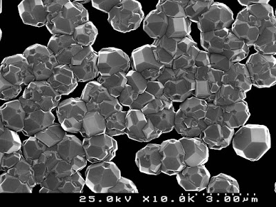
A new means of noninvasively tracking nanodiamonds with magnetic resonance imaging has been developed by researchers, opening up a host of new applications.


Nanodiamonds -- synthetic industrial diamonds only a few nanometers in size -- have recently attracted considerable attention because of the potential they offer for the targeted delivery of vaccines and cancer drugs and for other uses. Thus far, options for imaging nanodiamonds have been limited. Now a team of investigators based at the Athinoula A. Martinos Center for Biomedical Imaging at Massachusetts General Hospital has devised a means of tracking nanodiamonds noninvasively with magnetic resonance imaging (MRI), opening up a host of new applications. They report their findings in the online journal Nature Communications.
"With this study, we showed we could produce biomedically relevant MR images using nanodiamonds as the source of contrast in the images and that we could switch the contrast on and off at will," says David Waddington, lead author of the paper and a PhD student at the University of Sydney in Australia. Waddington is currently working with Matthew Rosen, PhD, in the Low-Field Imaging Laboratory at the Martinos Center. "With competing strategies, the nanodiamonds must be prepared externally and then injected into the body, where they can only be imaged for a few hours at most. However, as our technique is biocompatible, we can continue imaging for indefinite periods of time. This raises the possibility of tracking the delivery of nanodiamond-drug compounds for a variety of diseases and providing vital information on the efficacy of different treatment options."
Waddington began this work three years ago as part of a Fulbright Scholarship awarded early in his graduate work at the University of Sydney, where he is a member of a team led by study co-author David Reilly, PhD, in the new Sydney Nanoscience Hub -- the headquarters of the Australian Institute for Nanoscale Science and Technology, which launched last year. As part of the Reilly group, Waddington played a crucial role in early successes with nanodiamond imaging, including a 2015 paper in Nature Communications. He then sought to extend the potential of the approach by collaborating with Rosen at the Martinos Center and Ronald Walsworth, PhD, at Harvard University, also a co-author of the current study. Rosen's group is a world leader in the area of ultra-low-field magnetic resonance imaging, a technique that proved essential to the development of in vivo nanodiamond imaging.
Previously, the use of nanodiamond imaging in living systems was limited to regions accessible using optical fluorescence techniques. However, most potential diagnostic and therapeutic applications of nanoparticles, including tracking of complex disease processes like cancer, call for the use of MRI -- the gold standard for noninvasive, high-contrast, three-dimensional clinical imaging.
In the present study, the researchers show that they could achieve nanodiamond-enhanced MRI by taking advantage of a phenomenon known as the Overhauser effect to boost the inherently weak magnetic resonance signal of diamond through a process called hyperpolarization, in which nuclei are aligned inside a diamond so they create a signal detectable by an MRI scanner. The conventional approach to hyperpolarization uses solid-state physics techniques at cryogenic temperatures, but the signal boost doesn't last very long and is nearly gone by the time the nanoparticle compound is injected into the body. By combining the Overhauser effect with advances in ultra-low-field MRI coming out of the Martinos Center, the researchers were able to overcome this limitation -- thus paving the way for high-contrast in vivo nanodiamond imaging over indefinitely long periods of time.
High-performance ultra-low-field MRI is itself a relatively new technology, first reported in Scientific Reports in 2015 by Rosen and Martinos Center colleagues. "Thanks to innovative engineering, acquisition strategies and signal processing, the technology offers heretofore unattainable speed and resolution in the ultra-low-field MRI regime," says Rosen, director of the Low-Field Imaging Laboratory, an assistant professor of Radiology at Harvard Medical School and the senior author of the current paper. "And importantly, by removing the need for massive, cryogen-cooled superconducting magnets, it opens up a number of new opportunities, including the nanodiamond imaging technique we've just described."
The researchers have noted several possible applications for their new approach to nanodiamond-enhanced MRI. These include the accurate detection of lymph node tumors, which can aid in the treatment of metastatic prostate cancer, and exploring the permeability of the blood-brain barrier, which can play an important role in the management of ischemic stroke. Because it provides a measurable MR signal for periods of over a month, the technique could benefit applications such as monitoring the response to therapy.
Included in treatment monitoring are applications in the burgeoning field of personalized medicine. "The delivery of highly specific drugs is strongly correlated with successful patient outcomes," says Waddington, who was honored with the Journal of Magnetic Resonance Young Scientist Award at the 2016 Experimental NMR Conference in recognition of this work. "However, the response to such drugs often varies significantly on an individual basis. The ability to image and track the delivery of these nanodiamond-drug compounds would, therefore, be greatly advantageous to the development of personalized treatments."
The researchers continue to explore the potential of the technique and are now planning a detailed study of the approach in an animal model, while also investigating the behavior of different nanodiamond-drug complexes and imaging them with the new capability.